


Coronary Artery Disease
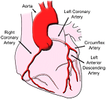
Coronary artery disease (CAD) affects almost 1.3 million Americans, making it the most common form of heart disease. CAD and its complications, like arrhythmia, angina pectoris, and heart attack (also called myocardial infarction), are the leading causes of death in the United States. CAD most often results from a condition known as atherosclerosis, which happens when a waxy substance forms inside the arteries that supply blood to your heart. This substance, called plaque, is made of cholesterol, fatty compounds, calcium, and a blood-clotting material l called fibrin. Doctors have found that there are 2 kinds of plaque: hard and soft.
Most people know about hard plaque and how it can cause a heart attack. If hard plaque builds up in the arteries that supply blood to your heart, the blood flow slows or stops. This decreases the amount of oxygen that gets to the heart, which can lead to a heart attack.
But doctors have now found that even though some heart attacks are caused by hard plaque, most heart attacks are caused by soft or vulnerable plaque.A vulnerable plaque is an inflamed part of an artery that can burst. This can lead to the formation of a blood clot, which can lead to heart attack.
What causes atherosclerosis?
Scientists think the disease starts when the very inner lining of the artery (the endothelium) is damaged. High blood pressure, high levels of cholesterol and triglycerides in the blood, and smoking are believed to lead to the development of plaque.
What are the symptoms?
Atherosclerosis may be present for years without causing symptoms. This slow disease process can begin in childhood. In some people, the condition can cause symptoms by the time they reach their 30s. In others, they do not have symptoms until they reach their 50s or 60s. But, as the blockage gets worse, the slowed blood supply to the heart may begin to cause something called angina pectoris, a Latin phrase that means, "strangling in the chest." Patients often say that angina is like a squeezing, suffocating, or burning feeling in their chest. The pain usually happens when the heart has an extra demand for blood, like during exercise or times of emotional stress.
Angina tends to start in the center of the chest but may move to your arm, neck, back, throat, or jaw. Some people say they feel numbness or a loss of sensation in their arms, shoulders, or wrists. An episode usually lasts no more than a few minutes and goes away with rest.
For certain patients with CAD, angina may not be present. Sometimes the lack of oxygen to the heart (called ischemia) does not cause any pain. In these cases, people are said to have silent ischemia.
How is CAD diagnosed?
Your doctor will take a medical history, ask about your symptoms, listen to your heart with a stethoscope, and perform certain tests, often including a chest x-ray. Here is a list of other tests that your doctor may order.
· A baseline electrocardiogram(ECG or EKG), which records your heart's electrical activity while you sit quietly. An exercise ECG, also known as a stress test, will show how your heart responds to increasing exercise. Both tests are designed to show if your heart is not working properly, most likely due to a lack of oxygen.
· An exercise thallium test, also called a nuclear stress test, which uses a radioactive substance that is injected into your bloodstream to show how blood flows through your arteries. Doctors can see if your heart muscle is damaged or dead, or if you have a serious narrowing in an artery. For people who cannot take an exercise test, medicines can be given that make your heart beat like it would if you were exercising.
· Echocardiography, which uses sound waves to produce an image of the heart to see how it is working.
· Coronary angiography, which is performed in the cardiac catheterization laboratory. After you are given medicine to relax you, dye is injected into your bloodstream to give doctors an x-ray "movie" of heart action and blood flow through your valves and arteries (called an angiogram). Doctors can see the number of blockages that you have and how serious those blockages are. Doctors often use this test to find out which treatment option may be best for you.
· Positron emission tomography(PET) scanning, which uses information about the energy of certain elements in your body to show whether parts of the heart muscle are alive and working. A PET scan can also show if your heart is getting enough blood in order to keep the muscle healthy.
How is CAD treated?
Medicines
A number of medicines can help relieve the angina pain that comes with CAD. People who have severe angina are often given a number of different medicines. Aspirin may also be given to patients with angina, because it decreases the chances of blood clots forming at the sites of the blockages.
· A medicine called nitroglycerin(nitro) can widen or dilate the arteries and improve blood flow to your heart. Nitro can be given through a skin patch, pills, an ointment, or a spray.
· Beta blockers "block" the chemical or hormonal messages sent to your heart. When you are under physical or emotional stress, your body sends signals to your heart to work harder. Beta-blockers block the effect these signals have on your heart, so they reduce the amount of oxygen your heart demands.
· Calcium channel blockers can help to keep your arteries open and reduce your blood pressure by relaxing the smooth muscle that surrounds the arteries in your body. The oxygen demand of the heart is also reduced by these medicines.
Transcatheter Interventions and Surgery
Because medicines cannot clear blocked arteries, a severely narrowed coronary artery may need more treatment to reduce the risk of a heart attack. Two major options are available: transcatheter interventions or coronary artery bypass surgery.
Both therapies have good track records among carefully selected patients. The decision to go with either option depends on how much narrowing there is, how many arteries are affected, the location of the narrowing, how much heart muscle is at risk, and individual patient factors, such as age and overall health.
Transcatheter Interventions
· Angioplasty, which opens narrowed arteries, is performed by interventional cardiologists. They use a long, thin tube called a catheter that has a small balloon on its tip. They inflate the balloon at the blockage site in the artery to flatten the plaque against the artery wall. Angioplasty is also called percutaneous transluminal coronary angioplasty (PTCA).
Here is how the procedure works. A thin wire is inserted into an artery in the leg and is guided to the site of narrowing in the coronary artery. The catheter is slipped over this guidewire and positioned at the blockage, where the balloon is inflated. After treatment, the wire, catheter, and balloon are removed. The hospital stay and recovery time for angioplasty is shorter than that of bypass. But about 35% of patients are at risk of more blockages in the treated area. This is called restenosis. Restenosis usually happens within 6 months after angioplasty.
· A stent procedure is used along with balloon angioplasty. It involves placing a mesh-like metal device into an artery at a site narrowed by plaque. The stent is mounted on a balloon-tipped catheter, threaded through an artery, and positioned at the blockage. The balloon is then inflated, opening the stent. Then, the catheter and deflated balloon are removed, leaving the stent in place. The opened stent keeps the vessel open and stops the artery from collapsing. Restenosis rates with this procedure are generally around 15% to 20%.
Because restenosis is a problem with the stent procedure, doctors have been trying to find ways to keep arteries with stents open. Some newer stents are covered with medicines that help keep the artery from closing up again. These are called coated stents or drug-eluting stents. In another stent procedure called brachytherapy, doctors deliver a low dose of radiation directly to the stent. It is thought that the radiation may shrink the tissue blocking the stent and stop it from growing back.
Surgery
Coronary artery bypass surgery means "bypassing" blood flow around one or more narrowed vessels. To do this, the surgeon removes a vein from the thigh (called the saphenous vein) or uses an artery from the upper part of the chest wall (called the internal mammary artery). Sometimes, an artery from another area of the body may be used. This surgically removed vessel is called a graft. The graft may be cut into sections for use in routing blood flow around blocked coronary arteries. After making an incision through the chest, the surgeon connects a graft at points above and below the blockage to restore blood flow.
· Minimally invasive coronary artery bypass is a less invasive bypass surgery technique. The incision is smaller, and the procedure may be done while the heart is still beating. This reduces the risk of complications. The procedure may reduce patient recovery time, which decreases cost. This operation is only used for patients whose blockages can be bypassed through this smaller incision and whose risk of complications is low.

Copyright ©1996-2012 Michigan Physicians Group. All Rights Reserved